
History
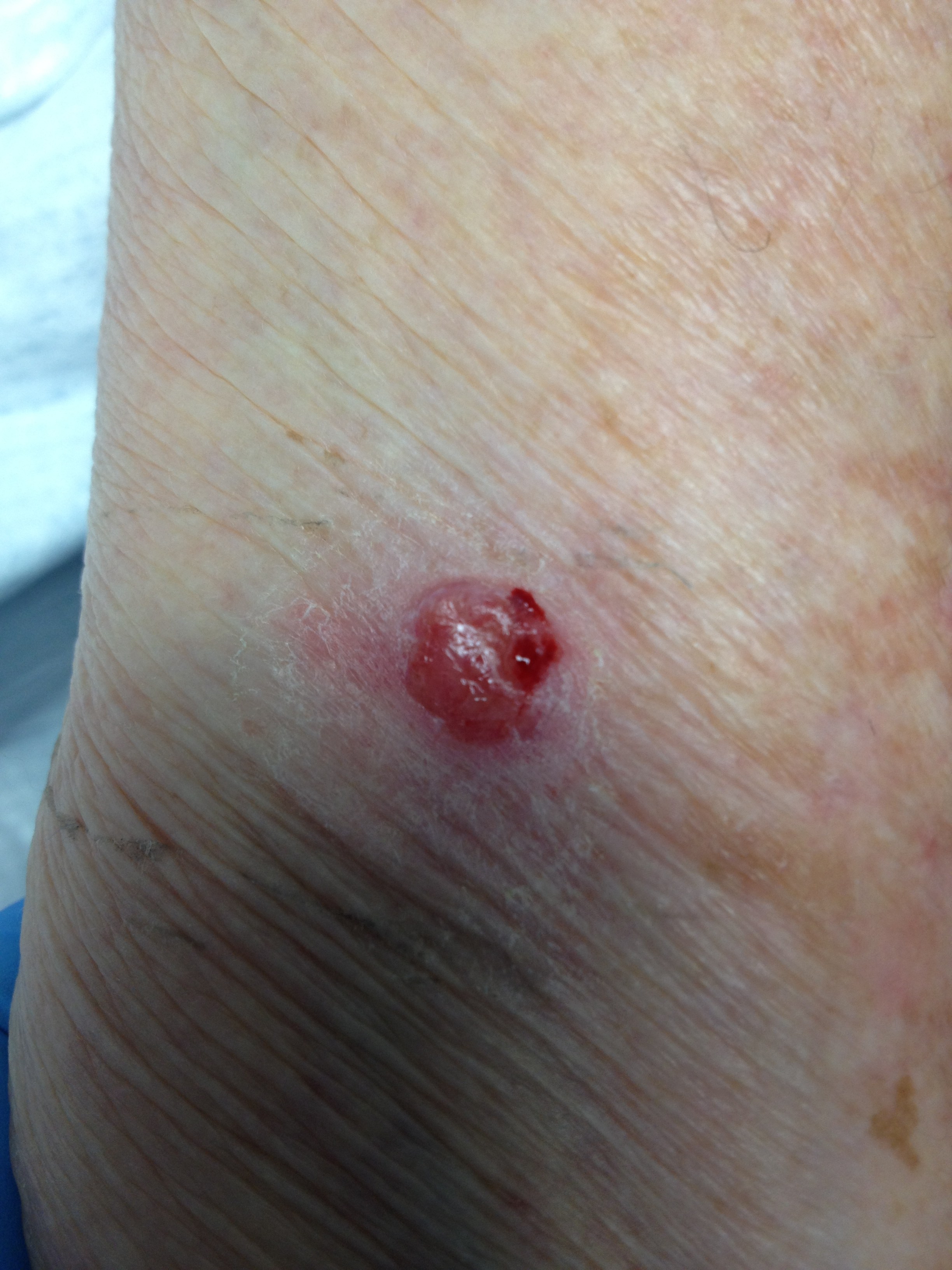
This patient is a 76 year old female who presented for the evaluation of a left forearm non-healing wound. She states it began back in June 2011 and as of March 2012 it had not healed. The inciting event was the placement of a screening PPD so she could enter an assisted-living facility. Since that time a nodule formed which she did not describe as painful. She did report it to be friable with a tendency to bleed easily. Treatment prior to coming to our facility was neosporin and topical hydrocortisone. Her medical history is positive for atrial fibrillation, hypertension and hyperlipidemia.
Wound characteristics
0.8cm x 0.7cm x 0.3 cm raised lesion. Very friable and bled easily to the touch.
Plan
Due to the long-standing nature and atypical appearance of this area it was anesthetized with xylocaine with epinephrine and a 3mm punch biopsy was performed. Punch biopsy was preferred over shave biopsy due to the history associated with this lesion. Since it was initiated by a PPD, I favored punch biopsy so I could see if there was some involvement of the dermal-epidermal junction. A shave biopsy might have missed this important data.
Diagnosis
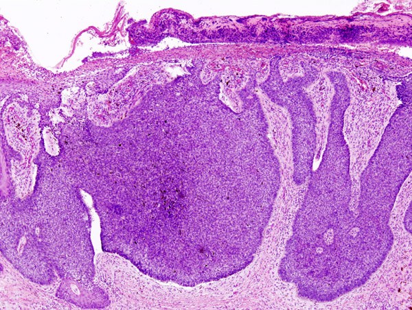
Basal cell carcinoma
Treatment
Patient was referred to surgery for excision of the mass.
Leave a Reply