
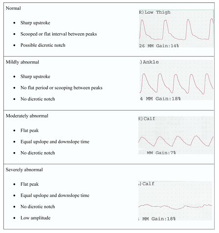
Waveform analysis including evaluation of the shape and amplitude of the wave forms is another helpful tool in the vascular assessment of the diabetic patient. Commonly ABI tests and segmental pressure tests are accompanied by their corresponding waveforms. These waveforms are interpreted based on standardized criteria relating to their anatomic location and severity of disease. The waveform is obtained by injecting a standardized volume of air into the pneumatic cuffs. The volume of air injected occludes venous circulation while not impacting arterial circulation. Changes occurring beneath the cuffs are then translated into pressures waves that are displayed as waveforms.
A normal waveform contains a sharp systolic upstroke, and sharp peak with a sharp and distinct downstroke and a visible dicrotic notch. A hemodynamically significant lesion will dissipate energy and change the contour of the recorded waveform. Below is a table of normal and abnormal pulse volume recording from the Journal of the American Society of Echocardiography as well as the full text article discussing other vascular ultrasound test.
Leave a Reply