
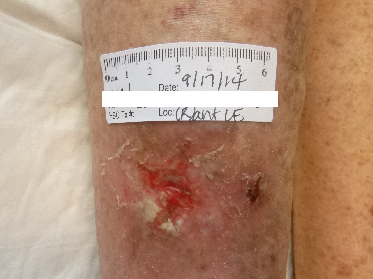

It is not uncommon to see a patient with sickle cell disease present to a wound care center with leg ulcerations. On first thought, one might postulate that these ulcers result from lack of oxygen delivery to the tissues due to mutated, sickle-shaped erythrocytes with reduced oxygen content. In actuality, it is long term use of hydroxyurea, the medication used to prevent sickle cell pain crises that is implicated in the development of these wounds.
Hydroxyurea is an antimetabolite used in the treatment of chronic myeloproliferative disorders. It works by inhibiting the S phase of DNA synthesis, and maintaining the cells in the G1 phase of the cell cycle. In sickle cell disease, it works by increasing the amount of hemoglobin F, which has a higher oxygen binding capacity than adult hemoglobin. It also results in the development of megaloblastic erythrocytes with increased water content, altered deformability, and reduced adherence to the endothelium. Cell size returns to normal within 24 hours of cessation of therapy. Adverse effects include bone marrow suppression, vascular ulcerations and gangrene, and erythrocyte abnormalities.
A retrospective study published in 1999 looked closely at the adverse effects of hydroxyurea in an attempt to elucidate the underlying pathophysiology behind the development of leg ulcers. 41 patients (mean age 67) with myeloproliferative disorders and no underlying vascular disease who underwent hydroxyurea therapy for a mean of 5 years developed leg ulcerations. 33 of these patients (80%) achieved full healing of their wounds upon cessation of the hydroxyurea therapy. The 8 remaining patients did experience some improvement and a reduction in the ulcer size. It was also noted that resuming hydroxyurea therapy resulted in recurrence of the leg ulcerations.
The study concluded that the pathophysiology behind ulcer development stems from the megaloblastic nature of the erythrocytes. The large red blood cells with altered deformability are unable to circulate optimally in the distal capillary beds, leading to cutaneous anoxia and subsequent ulceration. The fact that the erythrocytes become normocytic shortly after discontinuation of hydroxyurea is consistent with the clinical findings observed in this study.
This is just one example of a case where the best treatment might be simply removing an obstacle to the healing process. If the patient can tolerate it, hydroxyurea discontinuation is something to highly consider before ordering additional medications, labs and studies.
Contributing author Erin Fitzpatrick, MS3 Campbell University College of Osteopathic Medicine
References:
“Hydroxyurea: Drug Information.” UpToDate. Lexicomp, n.d. Web. 25 Nov. 2015. <http://www.uptodate.com.proxy.campbell.edu/contents/hydroxyurea-druginformation?source=search_result&search=hydroxyurea&selectedTitle=1~150>.
Sirieix, Marie-Emmanuelle, MD. “Leg Ulcers and Hydroxyurea.” JAMA Network. N.p., 1999. Web. 25 Nov.<http://archderm.jamanetwork.com/article.aspx?articleid=477918>.
Leave a Reply